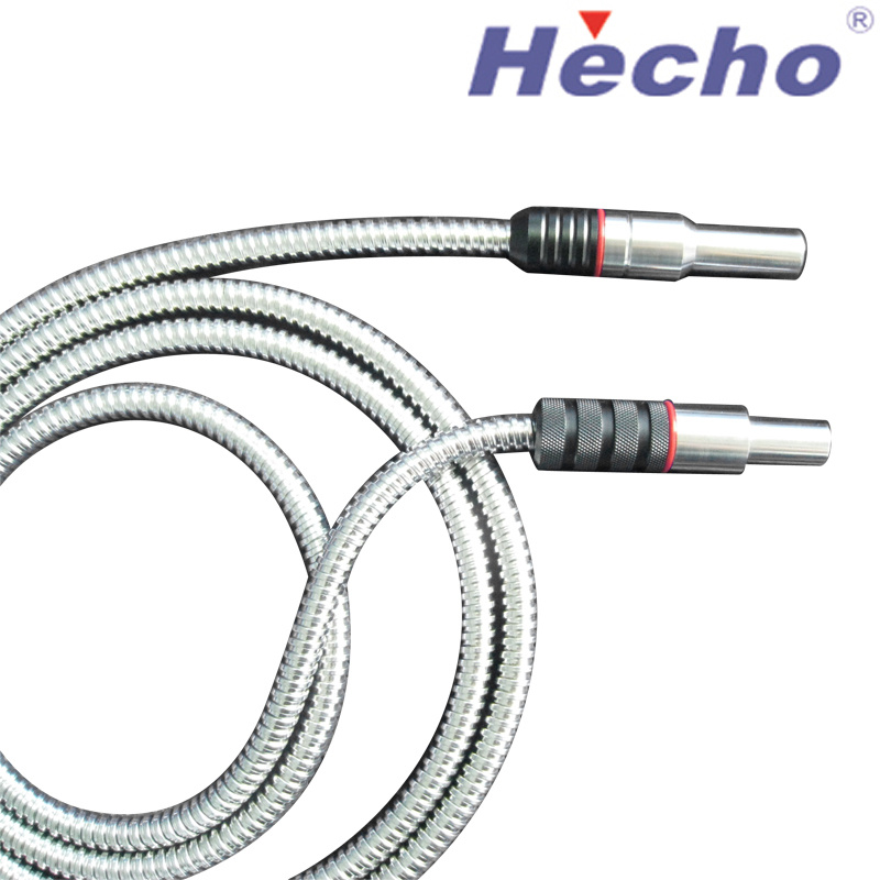

Bensing Section Mesh Applications
Bending section mesh is a delicate stack of stainless steel braids that are used to reduce friction and increase the bending strength of wire rope. It is available in various thicknesses and is easy to install.
Bensing section mesh can be applied in many types of bending applications. It increases the flexural strength of wire ropes and improves bending resistance of other curved components.
Endoscopes
Endoscopes are a type of medical instrument that is used to examine the gastrointestinal (GI) tract. These instruments are small, light and flexible and can be inserted into your mouth and moved down the esophagus, stomach and duodenum. The endoscope can also be used to remove small pieces of tissue, called biopsies, for examination in a lab.
The most common type of endoscope is the flexible video endoscope, which traces the GI tract with a camera at the tip. The endoscope’s image is viewed on a video monitor in the exam room. The provider uses the endoscope to look for any problems or perform procedures such as removing a cancerous tumor from the digestive tract with the help of a needle.
A 300-W xenon arc lamp in the endoscope’s light source generates the intense white, bright, high-intensity illumination needed for the imaging process. A quartz lens in the light guide at the distal tip of the endoscope collects and directs this illumination throughout the visual field. The light source is connected to the control section of the endoscope via a universal cord. A water container in the light source also contains water for cleaning and insufflation of the insertion tube during the procedure.
To supply the air, water and suction needed to inflate the insertion tube during the procedure, an air pump is located in the light source. This air pump forces a pipe protruding from the light source connector up the universal cord to the air/water valve on the control section of the endoscope (see Fig. 3.9). When the endoscopist releases the air/water valve, a vent hole in this valve allows the air to escape freely. This feature eliminates the risk of accidentally overinsufflating the patient’s GI tract, which could result in complications.
In addition to preventing overinsufflation, this system also prevents accidental ingestion of fluid during the procedure. A water bottle attached to the light source or cart is a convenient reservoir for storing a large volume of water during the procedure, which is used to flush away fluid from the insertion tube and objective lens.
Biopsies
Biopsies are a medical procedure that collects tissue or cells from an Bensing Section Mesh area of the body for examination in a laboratory. They are used to diagnose cancer and other diseases.
Most biopsies are performed to examine tissue in an abnormal area or lump. These are usually ordered when imaging tests can’t clearly identify the problem or if the abnormality is not responding to treatment.
A biopsy can be done using a needle, a scalpel, or a special vacuum-assisted device that uses suction to collect the cells. The type of biopsies your doctor performs will depend on where they are done and the type of cancer that is suspected.
Needle biopsy is a very reliable way to get samples of tissue. These samples can help your doctor determine whether a nodule is benign (non-cancerous) or malignant (cancerous).
When you go in for a needle biopsy, you will receive a local anesthetic to numb the area. You may also receive a sedative to help you relax.
Your doctor will make a small nick in the skin at the site of the nodule and use imaging guidance to guide the needle into the area. Once the needle is in place, it will advance to the site of the nodule and remove a sample of tissue for examination in the lab.
You might feel a slight pin prick or pressure as the needle enters the skin, but it will numb very quickly. You will need to stay still while the procedure is done.
The pain you feel after the procedure will vary depending on how much tissue was removed and what parts of the body were biopsied. Some people might experience a dull ache or slight discomfort for several days.
Some breast biopsies are done using a special device called a vacuum-assisted biopsy. A special machine sucks out the cells and sends them to the lab.
Other types of biopsies are done with a needle, or through an endoscope. These biopsies are typically done by surgeons, but they can also be done by other doctors in a hospital.
Endovascular Devices
Endovascular devices are used to treat aneurysms in the aorta. These devices are made up of a fabric tube framed with stainless steel self-expanding stents that are placed in the aorta and then expanded to seal off the aneurysm. This method is often more effective than open surgery because it reduces the risk of blood flowing into the aneurysm, but it can also lead to complications such as graft migration or stent buckling.
There are many different types of aortic endovascular grafts that have been developed over the years. Each device has a unique set of features, such as the stent material, graft fabric, fixation method, deployment mechanism, and flexibility. Most of these features have been improved since the first generation of aortic endografts, but there are still issues with their durability and compliance.
The underlying issue with aortic endografts is that the metal skeleton in the device has a very stiff and rigid design, which can result in a mismatch in physio-mechanical properties between the stented aorta and the native aorta. This mismatch can increase the workload and impedance on the heart, which leads to aortic regurgitation or rupture.
To combat this, several strategies have been implemented. One approach is to place hooks or anchors into the graft to embed it in the aortic wall. Another is to use radial force to fix the graft in place. These techniques are relatively easy to implement, but they can be very problematic when used on complex visceral anatomy.
Despite these challenges, endovascular aortic grafts have become an increasingly common treatment option for AAAs. They are effective, safe, and durable, but Bensing Section Mesh they do require lifelong follow-up to monitor for adverse events.
The latest generation of aortic endografts are made with stainless steel materials that are designed to improve the durability of the graft and reduce the likelihood of endoleaks. These devices include the AFX with Strata, AFX with Duraply, and AFX2 endovascular grafts from Endologix. The FDA has recommended that patients who have undergone implantation with any of these devices undergo yearly, lifelong imaging follow-up to monitor for Type III endoleaks.
Implants
Implants are used in a variety of Bensing Section Mesh applications, including breast reconstruction and revision surgery, rhinoplasty, nipple regeneration after mastectomy, reconstructive surgery for cleft palate, hernia applications and long bone fractures. The implants must be able to withstand compressing forces while being flexible and adaptable to the underlying tissue structures at the implant site. They should also possess mechanical properties that are predictable and easily controlled with sufficient strengths for the specific clinical need.
One solution to the problems mentioned above is to provide a resorbable polymeric medical implant, preferably in a three-dimensional form with a structure that allows for rapid tissue in-growth. This may be achieved through the use of materials such as polymers made from the monomers glycolide, lactide and all stereoisomers thereof, trimethylene carbonate, e-caprolactone, dioxanone or dioxepanone, or combinations of these.
The mesh material should be capable of bending without breaking and also have a pore homogeneity that is consistent throughout the entire implant structure. The porosity of the resorbable material should be higher than 70% so that it can withstand compression, but it should still be flexible enough to be adaptable to underlying tissue structures.
To control the bending strength of a mesh implant, it is necessary to provide it with a number of extra connections, i.e. threads or fibers that extend between and adjoin adjacent crests, as well as cuts along crests to reduce the bending strength of the resorbable mesh.
However, these extra connections can lead to a directional difference in the bending strength of the implant when a force is applied perpendicular to the crests, i.e. the bending strength is higher in the direction of the crests than when the force is applied parallel to them.
This directional difference in the bending strength of corrugated or waffled structures can be compensated by reducing the number of crests or providing a structure with a curved shape, for example by means of dedicated annealing processes. Another option is to give the resorbable material a rolled configuration, i.e. by forming the resorbable material into a spiral, which may be useful for implantation in abnormal bodily orifices such as fistulas but more specifically within such areas as rhinoplasty, nipple augmentation after mastectomy, reconstructive surgery for rhinoplasty, nipple, chin and hernia regeneration as well as long-bone fractures.